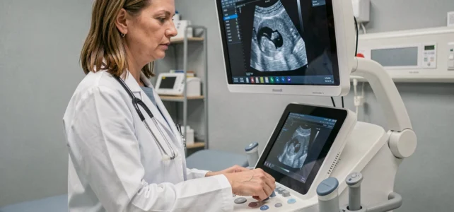
The image quality of an ultrasound machine depends less on the brand displayed on the chassis than on the architecture of the imaging engine and the choice of probes. Before comparing quotes, we recommend establishing a precise technical framework: required imaging modes, probe frequencies suitable for your practice, and connectivity constraints. Knowing how to choose a high-performance ultrasound machine starts with a clinical assessment grid, not a commercial catalog.
Imaging Engine and Signal Processing: The Foundation of Ultrasound Performance
The imaging engine (beamformer) determines spatial resolution, contrast resolution, and frame rate. A multi-channel digital beamformer offers a significantly higher number of firing lines per image compared to a retrofitted analog architecture. In practice, this results in a finer lateral resolution on deep structures, where entry-level devices lose definition.
To establish a rigorous selection framework, understanding how to choose a high-performance ultrasound machine involves comparing the specifications of the beamformer with the actual requirements of your specialty.
We observe that frame rate remains a neglected point in consumer comparisons. In cardiology or dynamic musculoskeletal ultrasound, an insufficient frame rate degrades real-time motion analysis. An engine capable of maintaining a high frame rate in simultaneous color Doppler mode provides a concrete advantage in daily practice.
Post-beamforming signal processing (adaptive filtering, speckle reduction, tissue harmonic imaging) follows. Tissue harmonic imaging improves contrast in patients with challenging morphology by exploiting the harmonic frequencies generated by the tissues themselves rather than the fundamental frequency. This mode should be included in the non-negotiable list of any specifications document.
Ultrasound Probes: Frequency, Bandwidth, and Compatibility
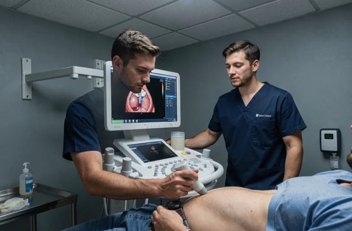
The probe is the link that directly determines diagnostic quality. A wideband probe covers an extended frequency spectrum, allowing a switch between superficial resolution and deep penetration without changing transducers. This point has a direct impact on workflow in consultations.
Three Categories of Probes to Master
- The low-frequency convex probe remains the reference for abdominal, pelvic, and obstetric exploration. Its typical bandwidth covers the frequencies necessary for sufficient penetration on corpulent patients.
- The high-frequency linear probe targets superficial structures: thyroid, peripheral vascular, musculoskeletal. Its axial resolution allows distinguishing structures of a few millimeters.
- The endocavitary probe (transvaginal or transrectal) imposes specific disinfection requirements. Recent studies remind us that a protocol aligned with the manufacturer’s instructions (IFU) remains necessary, even in outpatient settings.
A common mistake is to acquire an ultrasound machine with only one probe to limit the initial budget, only to find that actual clinical indications require a second transducer. Checking the compatibility of the proposed probe inventory with the unit before purchase avoids significant additional costs later.
Artificial Intelligence and Diagnostic Assistance in Ultrasound
AI embedded in recent ultrasound machines goes beyond simple image enhancement. A review published in August 2026 on thyroid ultrasound describes functions ranging from artifact suppression to automatic segmentation, as well as quantification and structured reporting. These tools reduce inter-operator variability, a recurring issue in ultrasound.
AI becomes a concrete differentiator in choosing an ultrasound machine, beyond raw image quality. In practice, automating biometrics (fetal measurements in obstetrics, bladder volume calculations) saves several minutes per exam. For a practice performing several dozen procedures per week, the cumulative savings are substantial.
We recommend testing AI functions in real conditions during the demonstration, not just on pre-recorded cases optimized by the manufacturer. Performance on varied morphologies and degraded acoustic windows reveals the actual robustness of the algorithm.
Cybersecurity of Connected Ultrasound Machines: A Now Technical Criterion
Modern ultrasound machines integrate network connectivity (DICOM, Wi-Fi, cloud) for archiving and sharing images. This connectivity exposes the device to the same vulnerabilities as any networked equipment. The FDA update published on February 3, 2026, reminds us that connected devices must integrate a post-market vulnerability plan and a SBOM (software bill of materials). In Europe, regulations are evolving in the same direction.
In practice, we recommend checking three points when purchasing:
- Does the manufacturer publish regular security updates, and for how long do they commit?
- Does the system allow for patient data encryption at rest and in transit?
- Is the SBOM available, listing the third-party software components integrated into the firmware?
An ultrasound machine whose software support cycle ends after a few years becomes a risk for the practice’s compliance. The duration of cybersecurity support conditions the actual lifespan of the investment.
Ultrasound Machine Format and Practice Context: Station, Portable, or Pocket
The choice of format directly stems from the mode of practice. A cart-based station offers a large screen, a full physical keyboard, and maximum computing power, but it remains confined to a dedicated room. A portable ultrasound machine combines image quality close to fixed stations thanks to miniaturization of components, with the ability to move between multiple sites.
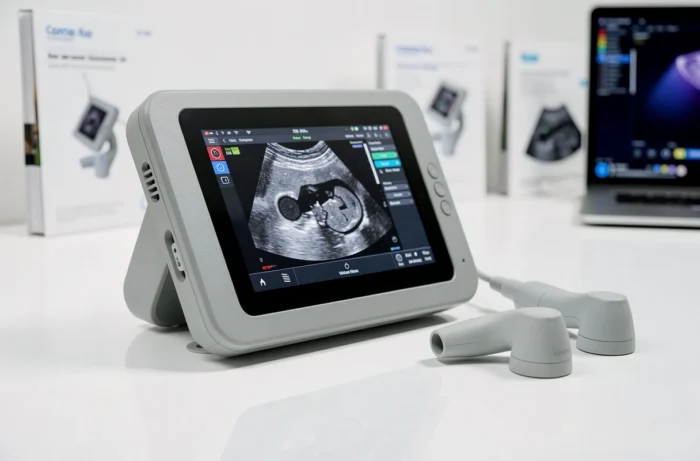
Pocket ultrasound machines connected to a smartphone or tablet cover triage echography and guidance of procedures. Their resolution lags for in-depth examinations, but their immediate availability at the patient’s bedside changes clinical practice.
The criterion not to overlook is the ergonomics of the keyboard and software interface. An ultrasound machine with poorly organized presets or navigation between modes requiring too many steps slows down each exam. Testing the interface in real consultation conditions remains the best way to evaluate this parameter, well before comparing technical sheets.
The choice of an ultrasound machine commits the practice for several years. Rather than reasoning solely on the acquisition price, incorporating the cost of additional probes, the duration of software support, and the availability of spare parts provides a more accurate view of the total budget. A machine that is cheaper to purchase but whose updates stop quickly costs more than a model that is better supported over time.